Department of Entomology
  • About
    • At a Glance
    • Welcome
    • Code of Conduct
    • Diversity, Equity, and Inclusion >
      • DEI Working Group
      • Resources
    • Departmental History
    • For Alumni
    • Support Entomology >
      • Steinhauer Scholarship Fund
    • Contact >
      • Directions
  • News
    • News
    • Seminar Blog
    • Seminar Schedule
    • Awards
  • People
    • Faculty
    • Post Docs
    • Students
    • Staff
    • Alumni
    • For PI/Faculty
    • Proposal Resources
  • Academics
    • Graduate >
      • Admissions
      • MS Degree Requirements
      • PhD Degree Requirements
      • Graduate Student Resources
      • Forms for Grad Students
      • Financial Assistance
      • Award & Funding Opportunities
      • Entomology Student Organization
    • Online Masters in Applied Entomology
    • Undergraduate >
      • Entomology Minor
      • Honors Program
  • Research
    • IPM & Biological Control of Agricultural, Urban & Forest Pests
    • Ecology, Conservation, Restoration, Climate Change >
      • Pollinator Science and Apiculture
    • Evolution, Systematics and Evo-Devo
    • Genetics & Genomics and Medical Entomology
  • Extension/Outreach
    • Educational Outreach
    • Insect Camp
    • Insect Drawings
    • Insect Identification
    • Pesticide Education and Assessment Program
    • Plant Diagnostic Laboratory (PDL)

NSF Graduate Research Fellowship awarded to former UMD Undergrad

3/31/2016

 
Alison Post (BS Biology 2014) received the NSF Graduate Research Fellowship. Alison is the former Lab Manager for the Lamp Lab and is now an ecology graduate student at Colorado State University. Check out the news here: https://cmns.umd.edu/news-events/features/3483. Congratulations Alison!

Plant Biotic Interactions: plants aren’t so defenseless after all

3/22/2016

 
Plants are sessile organisms and offer an abundance of food for hungry, relentless herbivores. To defend themselves, plants fight a silent chemical war with their herbivore foes. To do this, first plants must recognize mechanical damage inflicted by herbivores so that they can “decide” whether or not to make the costly investment of preparing and unleashing their chemical arsenal. Dr. Simon Zebelo from the University of Maryland, Eastern Shore has been researching how exactly plants do this. During his research, he discovered that oral secretions from beet armyworm caterpillars (Spodoptera exigua) combined with mechanical damage of plant tissue leads to induction of defense related responses in tomato plants. (Felton, 2008)

These oral secretions contain chemical compounds, called elicitors, which alert the plant to the herbivores presence. Where do these elicitors come from? The ventral eversible gland (VEG), which plays a role in caterpillar defense, seemed like a likely candidate because it was a secretory structure that came into contact with the plant while feeding. Using a heated pin, Dr. Simon and his co-workers destroyed the VEG and compared the ablated beet armyworm caterpillars to those with their VEG intact. They confirmed that the VEG secretions induce defenses in tomato plants (Solanum lycopersicum) that include increased expression of defense-related genes and emission of volatile organic compounds (VOCs). Thus, plants defend themselves by recognizing VEG originated elicitors and triggering their defense machineries.  

Next, Dr. Zebelo shifted his attention towards investigating a unique relationship between plants and plant-growth promoting rhizobacteria (PGPR). PGPR colonize the roots of plants and have been shown to promote plant growth through nitrogen fixation, hormone synthesis, nutrient uptake, and reduced plant disease. Dr. Zebelo and his co-workers are interested in how rhizobacteria mediate plant defense against insects. Dr. Zebelo and his colleagues were interested in seeing if PGPRs change the composition of VOCs. VOCs can deter herbivores and attract parasitic wasps which gladly parasitize the herbivores. They used a model system consisting of cotton (Gossypium hirsutum), beet armyworm (S. exigua), and a parasitoid wasp (Microplitis croceipes) to investigate the effect of treatment of cotton plants with single strain (INR7) or mixture of strains (Blend 8 and Blend 9) of PGPR (Bacillus spp.) on plant chemistry and plant-insect interactions. By inoculating cotton plants with strains of PGPR, collecting volatiles from collecting chambers (Fig 2), and then analyzing the quantity and quality of VOCs, it was became clear that PGPR cause the plants to produce more VOCs.

Following these results Dr. Zebelo and his co-workers tested whether the VOCs emitted by PGPR inoculated cotton plants affect the oviposition behavior of armyworm moths and the activity of parasitic wasps. Interestingly, beet armyworm moths laid fewer eggs on PGPR treated cotton plants and these PGPR treated plants were more attractive to parasitoids.

Gossypol is a secondary metabolite which has been linked with helping cotton plants protect themselves against herbivory by decreasing herbivore fitness. Dr. Zebelo and his co-workers questioned how PGPR affects the biosynthesis of gossypol and affect the feeding behavior of armyworm caterpillars. In a series of laboratory and greenhouse investigations, they demonstrated that PGPR treatment elicits the induction gossypol-related gene expression and this leads to increased levels of gossypol in cotton plants, which reduced herbivory by beet armyworm caterpillars.
​
This fascinating world of microbe-plant-insect interactions has important implications for agriculture. By exploring the largely unknown world of volatile ecology, we can identify new compounds that people can use to manage pests. However, Dr. Zebelo cautions against making too many generalizations at this early stage.  He reminds us that these interactions vary across plant, insect, and PGPR communities and are extremely complex, meriting additional research in this promising field.
           
References
 
Felton G. (2008) Caterpillar secretions and induced plant responses. In: Schaller, A., editor. Induced Plant Resistance to Herbivory. Netherlands: Springer. Chapter 18, 369–387.
 
Zebelo S., Piorkowski J., Disi J., & Fadamiro H. (2014). Secretions from the ventral eversible gland of Spodoptera exigua caterpillars activate defense-related genes and induce emission of volatile organic compounds in tomato, Solanum lycopersicum. BMC Plant Biology, 14(1), 140.
 
Zebelo S., Song Y., Kloepper J. W., & Fadamiro H. (2016). Rhizobacteria activates (+)-δ-cadinene synthase genes and induces systemic resistance in cotton against beet armyworm (Spodoptera exigua). Plant, Cell & Environment, 1-9.
 
 
About the Authors:
 
Hanna Kahl is a first year master’s student in Cerruti Hooks’ lab researching the effects of intercropping on insect feeding guilds.
 
Jonathan Wang is a PhD student in Raymond St. Leger’s lab. He is studying Drosophila immunity and fungal pathogenesis.
Picture
Figure 1 Anatomical diagram of Spodoptera exigua pointing out the ventral eversible gland (VEG)

Picture
Figure 2 Plants in volatile collection chambers

Not Your Friendly Neighborhood Spider

3/21/2016

 
An expert in her field as an investigative curator at the Smithsonian, Dr. Hannah Wood spends her time combing through leaf litter and examining tree trunks in Madagascar, Australia, New Zealand, South Africa and South America in pursuit of tiny and unusual predators.  She is investigating members of a small group of unique spiders known as the palpimanoids (Palpimanoidea). These arachnids may be small, (many measuring less than 10 mm in length) but these spiders are the stuff of nightmares for their prey. The highly modified cephalothorax (combination of the head and thorax) and elongated chelicerae (the spider’s jaws) make them excellent predators, well-adapted to their potentially dangerous and fast moving prey. Within the five known extant families of the Palpimanoidea, some are spider specialists, feeding only on other spiders, while some may subsist on small soil-dwelling arthropods, like Collembola. These spiders not only have unusual physical modifications, but also have uncommon methods of hunting, restricted distributions across the globe, low dispersal abilities, and can be traced back millions of years in the fossil record.  These qualities make them exciting prey for Dr. Wood, as she strives to learn how these tiny hunters developed their unique characteristics.

Palpimanoids have evolved over millions of years to use very unique methods to catch their prey. The two families of particular intrest to Dr. Wood are the Mecysmaucheniidae and the Archaeidae. In contrast to more typical spiders, these palpimanoids have elongated carapaces (the hardened cover over the cephalothorax) with a modified horizontal chelicerate muscle orientation, which is repsonible for operating their specialized, lengthened and especially mobile chelicerae.

One family that has captured the attention of Dr. Wood is the trap-jaw spiders (Family: Mecysmaucheniidae) which are native only to the cold mountain forests of New Zealand and southern South America. They use their jaws like a bear trap; they hold their long chelicerae out and open, which then snap shut when prey is close enough.  Dr. Wood’s dicovery of this unique mechanism is the first of it’s kind among arachnids. The speed at which some of these trap-jaw spiders are able to close their jaws is astonishing, particularly in a New Zealand species, which Dr. Wood has had difficultly observing even using a high-speed camera recording at 40,000 frames per second. Interestingly, this uncommon ability has evolved separately four times within this family. Furthermore, the trap-jaw spiders have developed a mechanism that allows them to store up large amounts of energy, which they then release at once to generate a greater force and speed to snap their chelicerae shut than would be possible with muscles alone. Dr. Wood is using CT scanning technology to get a closer look at the morphological mechanisms behind their incredible speed.                                                                                                                                                                                                                                  
In the mysterious forests of Madagascar, Australia and South Africa, Dr. Wood has investigated a close relative to the trap-jaw spider, known as the pelican spider (Family: Archaeidae). Pelican spiders have an even more an extreme carapace modification, which lends them the visual likeness to their namesake, pelicans, but you won’t see these minute predators diving for fish; they are spider specialists. These elaborate morphological adaptations have evolved to compensate for the muscles necessary to work their highly elongated chelicerae, which keep their potentially venomous prey at a distance when they strike. These extraordinary arachnids impale their prey with their venomous fangs positioned at the tip of their chelicerae, until their prey are immobile and no longer a threat, before lowering them to their mouthparts for consumption.   Dr. Wood has observed the diversification of these spiders on Madagascar and compared them to other restricted populations in Australia and South Africa to determine the evolutionary pressures acting on their diversification. This one of a kind research is not only interesting, but improves our understanding of evolution and speciation.
 
About the Authors:
Jackie Hoban is a first-year master’s student in Paula Shrewsbury’s lab studying emerald ash borer biological control.
Jen Jones is a first-year PhD student in Bill Lamp’s lab studying mosquitoes.

Picture
Figure 1 Chelicerae of a trap-jaw spider (Family Mecysmaucheniidae)

Picture
Figure 2 An example of the morphological anomaly of the pelican spiders (Family Archaeidae)

How the honey bee crisis is affecting California's almond growers 

3/16/2016

 
Entomology Graduate Students Nathalie Steinhauer and Meghan McConnell Collect Bees from California Almond Farm for USDA Testing – Los Angeles Times

Check out the full article here: 
goo.gl/6hSFZp
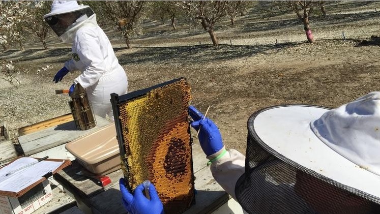
Picture
(Robin Abcarian / Los Angeles Times)

FDA says test of genetically modified mosquitoes is safe

3/16/2016

 
Entomology’s David O’Brochta Comments on the Testing of Genetically Modified Mosquitoes – NBC News

Check out the full article here: 
goo.gl/SnlvgQ
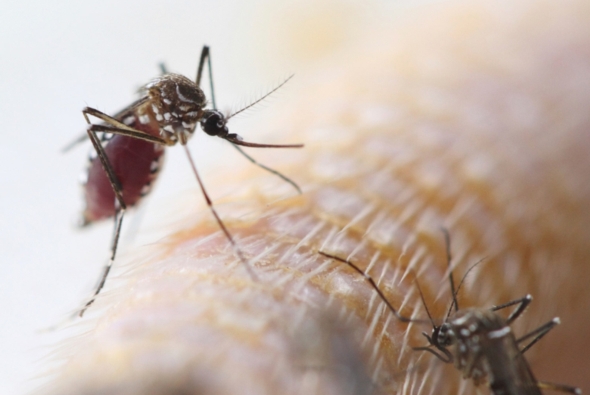
​

Picture
©iStock

Dr. Dilip Venugopal's recent PLoS paper featured on entomologytoday

3/10/2016

 
One of our recently graduated students Dr. Dilip Venugopal (now a AAAS fellow) has coauthored a paper related to brown- marmorated Stink Bugs in PLOS ONE. Check it out at goo.gl/CvAZ2c . You can also read his views here: goo.gl/1ZyLCf
Picture
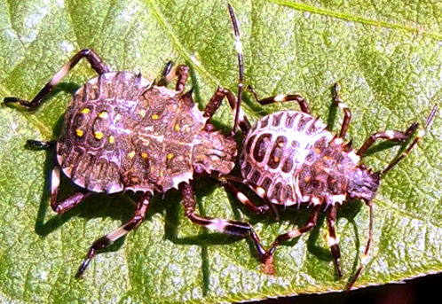
Photo by: Dr Dilip Venugopal

Viruses and Mites and Black Bees, Oh My!

3/9/2016

 
Picture
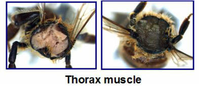
Figure 1. the thorax muscle tissue of a healthy bee (left) compared to that of a "Black Bee"
Dr. Humberto Boncristiani is a post-doc in the vanEngelsdorp bee lab at the University of Maryland.  He is currently studying a variety of honey bee viruses, with a particular focus on picorna-like viruses.  His interest in honey bees came from his father, who keeps bees in Brazil, and his virology background comes from his work on human viruses at the University of Sao Paulo.  The inspiration for his transition from humans to honey bees came in 2007 when Diana Cox-Foster published a paper documenting an association between Israeli acute paralysis virus (IAPV) and colony collapse disorder (CCD).  IAPV just so happened to belong to the same group of viruses that Humberto was studying in humans.  This prompted him to make the jump to studying honey bee viruses.
Picornaviruses have a wide range of hosts, from honey bees to humans.  They are of particular interest because of the manner in which they replicate.  Picornaviruses have an Internal Ribosome Entry Site (IRES) analogous to that of their host’s mRNA.  The IRES is a complex part of the viral RNA genome used to confuse the cellular machinery and inciting the ribosome to start translation of the virus proteins.  In order to prevent the cell from producing its own proteins, the picornavirus cleaves an important protein from the cellular mRNA making it unrecognizable to the ribosome (Boncristiani et al., 2009).  Finally, the picornavirus uses the cell’s ribosomes to replicate itself by the thousands.  The virus transcriptase lacks the ability to proofread itself, leading to higher mutational rates in the copies it makes.  This results in a cloud of genetically diverse viruses, most of which can cause physical symptoms and infect other cells.  The incredible genetic diversity in these viruses makes them dangerous pathogens that hosts have a difficult time evolving resistance to, which is why they have such a profound effect on bee colonies.
Some of Humberto’s current work is based on a phenomenon that has come to be known as “The Black Muscle Bees.”  These bees were discovered after processing a set of USDA-APHIS National Honey Bee Survey samples of adult honey bees.  The samples of bees were crushed in water to prepare a solution that could be analyzed for Nosema sp., a fungal pathogen that is commonly detected in honey bee colonies across the country.  The solution turned a dark shade of black, as opposed to the normal brown.   This unusual observation warranted further investigation, so more bees were collected from multiple colonies in the same yard of the original “Black Bees.” Upon dissecting these bees it was discovered that the tissue inside of symptomatic bees was entirely black when compared to the standard pink tissue of healthy bees.    It is thought that this darker color is caused by an increase in pigmentation or melanin formation. The melanin formation process is induced by a serine protease cascade involving the enzyme prophenyloxidase (PPO).  The symptomatic “Black Bees” have a high level of PPO compared to asymptomatic bees from the same apiary. These bees were screened for other viruses and it was discovered that Deformed Wing Virus (DWV), a picornavirus, was more prevalent in the colonies with symptomatic bees when compared to healthy bees from the same yard. Additionally, the genetic diversity of these DWV strains was much higher in the colonies containing “Black Bees”.  DWV is a virus transmitted by Varroa destructor, a parasitic mite that was introduced to the United States in the 1980s.  Varroa has since been documented as a vector of a variety of honey bee viruses.  It transmits viruses directly into the haemolymph, the insect equivalent of blood, of the bee as the mite feeds.  This mode of inoculation bypasses the typical GI detoxification pathway of the honey bee.  Varroa mites also provide these viruses with an additional place to replicate and diversify, ensuring that Dr. Boncristiani will have plenty of work to stay busy in his new field of honey bee virology. 
 
References:
Boncristiani, H., Criado, M.F., Arruda, E., (2009). Respiratory Viruses. In M. Schaechter (Ed.), Encyclopedia of Microbiology (p. 4600). Academic Press.
Cox-Foster, D. L.; Conlan, S.; Holmes, E. C.; Palacios, G.; Evans, J. D.; Moran, N. A.; Quan, P.-L.; Briese, T.; Hornig, M.; Geiser, D. M.; Martinson, V.; vanEngelsdorp, D.; Kalkstein, A. L.; Drysdale, A.; Hui, J.; Zhai, J.; Cui, L.; Hutchison, S. K.; Simons, J. F.; Egholm, M.; Pettis, J. S.; Lipkin, W. I. (2009). A Metagenomic Survey of Microbes in Honey Bee Colony Collapse Disorder. Science, 318, 283–287.
           
Blog post written by:
Olivia Bernauer is a first year Master’s student in Dennis vanEngelsdorp’s bee lab working with wild, native bees. Olivia is currently working with volunteers to monitor the floral preference of Maryland’s native pollinators. 
 
Andrew Garavito is a Master’s student in Dennis vanEngelsdorp’s Lab.  He is studying honey bees, with a focus on the diversity of pollen types brought in by foragers, and the effects of different pollen diets on bee health.
 

    Categories

    All
    Awards
    Colloquium
    Faculty Spotlight
    Fall 2013 Colloquium
    Fall 2014 Colloquium
    Fall 2015 Colloquium
    Fall 2016 Colloquium
    Featured
    Innovation
    News
    Publications
    Science Projects
    SESYNC
    Spring 2014 Colloquium
    Spring 2015 Colloquium
    Spring 2016 Colloquium
    Talks
    Undergraduate

    Archives

    April 2025
    March 2025
    February 2025
    December 2024
    November 2024
    October 2024
    June 2024
    May 2024
    April 2024
    March 2024
    December 2023
    November 2023
    October 2023
    September 2023
    August 2023
    July 2023
    June 2023
    May 2023
    April 2023
    March 2023
    February 2023
    January 2023
    December 2022
    November 2022
    October 2022
    September 2022
    August 2022
    July 2022
    May 2022
    April 2022
    March 2022
    February 2022
    January 2022
    December 2021
    November 2021
    October 2021
    September 2021
    August 2021
    July 2021
    June 2021
    May 2021
    April 2021
    March 2021
    February 2021
    January 2021
    December 2020
    November 2020
    October 2020
    September 2020
    August 2020
    July 2020
    June 2020
    May 2020
    April 2020
    March 2020
    February 2020
    December 2019
    November 2019
    October 2019
    September 2019
    August 2019
    July 2019
    June 2019
    May 2019
    April 2019
    March 2019
    February 2019
    January 2019
    December 2018
    November 2018
    October 2018
    September 2018
    August 2018
    July 2018
    June 2018
    May 2018
    April 2018
    March 2018
    February 2018
    December 2017
    November 2017
    October 2017
    September 2017
    June 2017
    May 2017
    April 2017
    March 2017
    February 2017
    January 2017
    December 2016
    November 2016
    October 2016
    September 2016
    August 2016
    June 2016
    May 2016
    April 2016
    March 2016
    February 2016
    January 2016
    December 2015
    November 2015
    October 2015
    September 2015
    August 2015
    July 2015
    June 2015
    May 2015
    April 2015
    March 2015
    February 2015
    January 2015
    December 2014
    November 2014
    October 2014
    September 2014
    August 2014
    June 2014
    May 2014
    April 2014
    March 2014
    February 2014
    January 2014
    December 2013
    November 2013
    October 2013
    September 2013

    RSS Feed

Picture
Picture
Picture
Department of Entomology 
University of Maryland 
4112 Plant Sciences Building 
College Park, MD 20742-4454
USA

Telephone: 301.405.3911 
Fax: 301.314.9290
Picture
Web Accessibility
  • About
    • At a Glance
    • Welcome
    • Code of Conduct
    • Diversity, Equity, and Inclusion >
      • DEI Working Group
      • Resources
    • Departmental History
    • For Alumni
    • Support Entomology >
      • Steinhauer Scholarship Fund
    • Contact >
      • Directions
  • News
    • News
    • Seminar Blog
    • Seminar Schedule
    • Awards
  • People
    • Faculty
    • Post Docs
    • Students
    • Staff
    • Alumni
    • For PI/Faculty
    • Proposal Resources
  • Academics
    • Graduate >
      • Admissions
      • MS Degree Requirements
      • PhD Degree Requirements
      • Graduate Student Resources
      • Forms for Grad Students
      • Financial Assistance
      • Award & Funding Opportunities
      • Entomology Student Organization
    • Online Masters in Applied Entomology
    • Undergraduate >
      • Entomology Minor
      • Honors Program
  • Research
    • IPM & Biological Control of Agricultural, Urban & Forest Pests
    • Ecology, Conservation, Restoration, Climate Change >
      • Pollinator Science and Apiculture
    • Evolution, Systematics and Evo-Devo
    • Genetics & Genomics and Medical Entomology
  • Extension/Outreach
    • Educational Outreach
    • Insect Camp
    • Insect Drawings
    • Insect Identification
    • Pesticide Education and Assessment Program
    • Plant Diagnostic Laboratory (PDL)