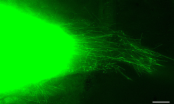
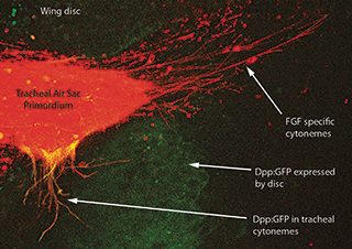
The Cytoneme Connection: Challenging our Knowledge of Paracrine SignalingAs we learned in colloquium earlier this semester, even the smallest arthropods have immense diversity in both body plan and development. According to Dr. Sougata Roy, a big contributing factor to this diversity is the ability of cells to communicate and induce changes in other cells. A major research interest of Dr. Roy’s is the function of cell structures called cytonemes in the development of Drosophila larvae. Cytonemes are well-known to be present during larval insect development and have been for quite some time, but until fairly recently, no one had researched their function. Belonging to a class of cellular protuberances called filipodia, cytonemes are filamentous structures known to be present in wing and eye imaginal disks in Drosophila and in the ovaries and embryos of several other invertebrates. Dr. Roy’s research suggests that these visually strange projections of the cell membrane play an integral role in paracrine signaling. Paracrine signaling is generally known to involve releasing signal molecules directly into the extracellular fluid, which reach target cells slowly through diffusion. Dr. Roy believed that cytonemes may somehow facilitate this process, allowing the molecules to better home in on their target cells. Armed with countless Drosophila and red and green fluorescent biomarkers, he conducted several experiments to see if this could be the case. Post by: Becca Wilson and Samuel Ramsey In one particularly illuminating experiment, Dr. Roy looked at the connections between cytonemes of cells in the wing discand tracheal system. He wanted to investigate whether the cytonemes needed to be in contact with each other for the signaling molecules to transfer. Cells of each type were marked with one part of a green fluorescent protein (GFP) that would fluoresce only at the contact point between the cells. The resulting images looked much like that scene from E.T., with a glowing light formed between the two spindle-like structures. Until this discovery only nerve cells were known to communicate this way. Now knowing that cytonemes of different cells did contact each other (sometimes over distances of 50 to 100 cells), Dr. Roy found a method to disable the distal molecule of the cytonemes. This did not impact the structure or number of the cytonemes, but they would no longer be able to form contact points. The resulting images were suddenly less colorful than usual, he concluded that disabling the connection of cytonemes resulted in no cell to cell communication. Through further research he was also able to determine that Drosophila cells have specific cytonemes for the transport of Fibroblast growth factors (FGF’s) and other signaling biomolecules integral to proper tissue differentiation. Having found that cytonemes are a surprisingly important structure in the case of FGF transfer in Drosophila, Dr. Roy hopes in the future to determine how cytonemes recognize their target cells. And, perhaps more importantly, if this is a universal mechanism or just a strange quirk to our favorite, markedly derived fruit fly. You can read more about Dr. Roy's research with cytoneme communication in the following papers: Cytoneme-mediated contact-dependent transport of the Drosophila Decapentaplegic signaling protein and Communicating by touch – neurons are not alone. About Becca Wilson and Samuel Ramsey:
Becca Wilson is a first year master's student in Bill Lamp’s lab. She is currently studying the distribution patterns of nuisance black flies in Washington County, Maryland. Samuel Ramsey is a 3rd year PhD student in the Shrewsbury lab currently studying competition in egg parasitoids of the brown marmorated stink bug. Comments are closed.
|
Categories
All
Archives
June 2024
|
Department of Entomology
University of Maryland
4112 Plant Sciences Building
College Park, MD 20742-4454
USA
Telephone: 301.405.3911
Fax: 301.314.9290
University of Maryland
4112 Plant Sciences Building
College Park, MD 20742-4454
USA
Telephone: 301.405.3911
Fax: 301.314.9290


 RSS Feed


