Department of Entomology
  • About
    • At a Glance
    • Welcome
    • Code of Conduct
    • Diversity, Equity, and Inclusion >
      • DEI Working Group
      • Resources
    • Departmental History
    • For Alumni
    • Support Entomology >
      • Steinhauer Scholarship Fund
    • Contact >
      • Directions
  • News
    • News
    • Seminar Blog
    • Seminar Schedule
    • Awards
  • People
    • Faculty
    • Post Docs
    • Students
    • Staff
    • Alumni
    • For PI/Faculty
    • Proposal Resources
  • Academics
    • Graduate >
      • Admissions
      • MS Degree Requirements
      • PhD Degree Requirements
      • Graduate Student Resources
      • Forms for Grad Students
      • Financial Assistance
      • Award & Funding Opportunities
      • Entomology Student Organization
    • Online Masters in Applied Entomology
    • Undergraduate >
      • Entomology Minor
      • Honors Program
  • Research
    • IPM & Biological Control of Agricultural, Urban & Forest Pests
    • Ecology, Conservation, Restoration, Climate Change >
      • Pollinator Science and Apiculture
    • Evolution, Systematics and Evo-Devo
    • Genetics & Genomics and Medical Entomology
  • Extension/Outreach
    • Educational Outreach
    • Insect Camp
    • Insect Drawings
    • Insect Identification
    • Pesticide Education and Assessment Program
    • Plant Diagnostic Laboratory (PDL)

[Seminar Blog] Reaching maturity? Malaria parasites’ developmental journey through the mosquito

11/4/2021

 
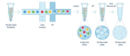
Written by PhD students Arielle Arsenault-Benoit & Minh Le

​Malaria is a mosquito-borne illness that has plagued human civilization for millennia (Cox 2010), and continues to infect many people, with over 200 million documented cases in 2019 (World Health Organization, 2020). Preventative interventions and treatments are available for those that can access them, yet according to the WHO, there are 400,000 annual deaths worldwide, and nearly 70% of those are children under 5 years old. Among those interventions are insecticide treated bed nets, residual insecticide spraying, preventative drugs, and treatment with combined drug therapy. Effective prevention and treatment efforts, plus advances in diagnostic testing, have greatly reduced both case numbers and mortality rates, but the development of resistance, including insecticide resistance in the mosquito vector population and drug resistance in the pathogen population present new challenges. A greater understanding of the underlying biology of the pathogen may allow for improved and more targeted interventions in the future. 
At our recent Department of Entomology colloquium, Dr. Haikel Bogale, a Post-Doctoral Fellow at the Institute for Genome Sciences, University of Maryland School of Medicine, discussed his work on the role of differential gene expression on the maturity of malaria parasites at different time points and at different locations within the mosquito body.
 
Malaria is caused by several species of unicellular parasites in the genus Plasmodium. These parasites have a complex life cycle, in which they undergo development, multiplication, and reproduction within the mammalian host and the mosquito vector. A female mosquito picks up the parasite from an infected host’s blood (i.e. human), then, while also going through its developmental stages, the parasite must enter the mosquito midgut via the blood meal, escape the mosquito’s immune system, break out of the midgut and into the body cavity, and travel through the hemolymph – analogous to blood in insects– to the mosquito’s salivary glands. Once in the salivary glands, the infectious stage of Plasmodium, called sporozoite, is transmitted to a mammalian host to cause disease and perpetuate the cycle. In an infected mosquito, thousands of sporozoites reach the salivary glands, but only a handful of those are injected to the mammalian skin. (For more detailed information about the life cycle of Plasmodium parasites, visit the CDC Malaria website.)
 
 
During an organism’s life cycle, different genes are expressed at different times, locations, and quantities in order to perform vital functions. In other words, individual genes are like different tools that an organism can retrieve and use to address different environmental pressures that they experience throughout their lifespan. The same concept holds true for malaria-causing Plasmodium parasites. Dr. Bogale leverages this notion to study how gene expression of Plasmodium parasites differs between multiple species’ sporozoites and between different anatomical locations in the mosquito where sporozoites reside.
 
When a gene is expressed, a class of molecules known as RNA is produced with a unique sequence. Reading and analyzing the RNA sequence profile of the sporozoites at certain stages can give us an idea of which genes are expressed at a particular stage and how much of that gene is being expressed. The traditional technique used to read the sporozoite RNA sequence is known as bulk RNA sequencing, which generates the RNA profile of a population of sporozoite cells. This technique, however, only produces a big picture of the population’s RNA profile and masks the minor variations between each sporozoite cell in that population (Chen et al., 2019). Dr. Bogale was among the first to employ an alternative technique called single cell RNA sequencing (scRNA-seq) to study Plasmodium sporozoites collected from multiple anatomical sites in the mosquito. As its name suggests, scRNA-seq makes it possible to sequence an individual sporozoite’s RNA profile within a sample containing thousands and thousands of sporozoites. This technique works by separating cells in individual oil droplets and allowing for the capture of information from messenger RNA (mRNA) of each cell (Figure 1), ultimately highlighting the differences in gene expression among all sporozoite cells within a given sample, a feat that is not possible with bulk RNA sequencing.
Workflow diagram for scRNA-seq.
Figure 1. Workflow diagram for scRNA-seq. Individual cells are enveloped in oil droplets containing a bead with a unique barcode. Barcodes are annealed onto the cells so data from individual single cells can be identified. From 10X Genomics. 2019. https://www.10xg
​Dr. Bogale’s scientific discoveries are two-fold. Firstly, he implemented scRNA-seq on the sporozoites of three different species of Plasmodium that cause malaria.  As a result, he was able to identify over 50 differentially expressed genes among these three species in the salivary gland and denote their functions (Bogale et al. 2021). For example, some genes that function in inhibition of protein degradation and translation inhibition are differentially expressed among three species of Plasmodium, demonstrating that there may be species-specific candidate genes to explore further.
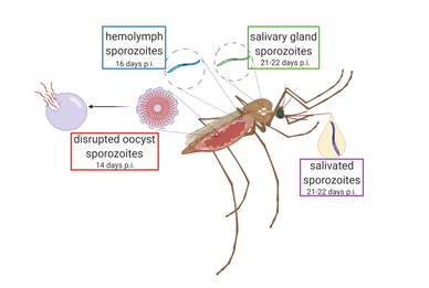
Secondly, Dr. Bogale’s group used scRNA-seq on sporozoites of a single species at four snapshots of their lifespan inside of the mosquito; from when they are just released from the oocyst, to when they are traveling in the mosquito’s hemolymph, when they inhabit the mosquito’s salivary glands, and then finally after they have been expelled from the mosquito’s proboscis by forced salivation (Figure 2). This allowed researchers to determine whether sporozoites within the same area of the mosquito are in the same stage of development and express the same genes. Researchers found RNA profiles from sporozoites that were collected from different mosquito anatomical sites can be similar (e.g., sporozoites from oocysts vs salivary glands), suggesting sporozoite maturation is not solely dependent on mosquito environment. However, they also identified over 50 differentially expressed genes across different sporozoite’s developmental stages (correlating with different anatomical residence). Lastly, they report an upregulation of components necessary for producing protein (ribosomal RNA) after salivation of sporozoites by the mosquito, in contrast to when it resides in the salivary gland. This could signify the transition from the sporozoite’s quiescence phase, where the parasite “sleeps” inside of the mosquito’s salivary gland, to a more active stage when it is trying to assemble the machinery needed to invade mammalian host cells.
Anatomical sites where sporozoites were harvested for this analysis
Figure 2. Anatomical sites where sporozoites were harvested for this analysis, with the respective timepoints when they were collected (days post infection).Sporozoites enter the hemolymph after oocyst mature and rupture, releasing them, around 14 days post-infection.From Bogale et al. 2021.
Researchers and public health professionals have taken numerous approaches to reduce the global burden of malaria, including drug therapies, vector management strategies, and most recently, the approval of a promising vaccine. Yet, new technologies and efforts are required to sustain this progress. Dr. Bogale’s research leading to a stronger understanding of genes and their functions involved in sporozoite maturity may allow researchers to target ways to suppress those genes or interrupt the machinery required to carry out the functions, potentially within the bodies of genetically modified mosquito vectors.
References:
 
Bogale, H.N., Pascini, T.V., Kanatani, S., Sá, J.M., Wellems, T.E., Sinnis, P., Vega-Rodríguez, J. and Serre, D., 2021. Transcriptional heterogeneity and tightly regulated changes in gene expression during Plasmodium berghei sporozoite development. Proceedings of the National Academy of Sciences, 118(10).
 
Chen, G., Ning B., Shi, T., 2019. Single-cell RNA-seq technologies and related computational data analysis. Frontiers in Genetics, 10: 317.
 
Cox, F.E., 2010. History of the discovery of the malaria parasites and their vectors. Parasites Vectors 3(5).
 
World Health Organization, 2020. World malaria report 2020: 20 years of global progress and challenges. In World malaria report 2020: 20 years of global progress and challenges.
 
Authors:
Arielle Arsenault-Benoit is a PhD student in the Fritz Lab. She studies the community composition and landscape genomics of Culex mosquitoes, vectors of West Nile virus, across a heterogeneous landscape. You can find her on twitter @BenoitArielle.
 
Minh Le is a PhD student in the Pick Lab. He studies the genetic networks involved in insect segmentation, specifically on hemipteran insects.

Comments are closed.

    Categories

    All
    Awards
    Colloquium
    Faculty Spotlight
    Fall 2013 Colloquium
    Fall 2014 Colloquium
    Fall 2015 Colloquium
    Fall 2016 Colloquium
    Featured
    Innovation
    News
    Publications
    Science Projects
    SESYNC
    Spring 2014 Colloquium
    Spring 2015 Colloquium
    Spring 2016 Colloquium
    Talks
    Undergraduate

    Archives

    April 2025
    March 2025
    February 2025
    December 2024
    November 2024
    October 2024
    June 2024
    May 2024
    April 2024
    March 2024
    December 2023
    November 2023
    October 2023
    September 2023
    August 2023
    July 2023
    June 2023
    May 2023
    April 2023
    March 2023
    February 2023
    January 2023
    December 2022
    November 2022
    October 2022
    September 2022
    August 2022
    July 2022
    May 2022
    April 2022
    March 2022
    February 2022
    January 2022
    December 2021
    November 2021
    October 2021
    September 2021
    August 2021
    July 2021
    June 2021
    May 2021
    April 2021
    March 2021
    February 2021
    January 2021
    December 2020
    November 2020
    October 2020
    September 2020
    August 2020
    July 2020
    June 2020
    May 2020
    April 2020
    March 2020
    February 2020
    December 2019
    November 2019
    October 2019
    September 2019
    August 2019
    July 2019
    June 2019
    May 2019
    April 2019
    March 2019
    February 2019
    January 2019
    December 2018
    November 2018
    October 2018
    September 2018
    August 2018
    July 2018
    June 2018
    May 2018
    April 2018
    March 2018
    February 2018
    December 2017
    November 2017
    October 2017
    September 2017
    June 2017
    May 2017
    April 2017
    March 2017
    February 2017
    January 2017
    December 2016
    November 2016
    October 2016
    September 2016
    August 2016
    June 2016
    May 2016
    April 2016
    March 2016
    February 2016
    January 2016
    December 2015
    November 2015
    October 2015
    September 2015
    August 2015
    July 2015
    June 2015
    May 2015
    April 2015
    March 2015
    February 2015
    January 2015
    December 2014
    November 2014
    October 2014
    September 2014
    August 2014
    June 2014
    May 2014
    April 2014
    March 2014
    February 2014
    January 2014
    December 2013
    November 2013
    October 2013
    September 2013

    RSS Feed

Picture
Picture
Picture
Department of Entomology 
University of Maryland 
4112 Plant Sciences Building 
College Park, MD 20742-4454
USA

Telephone: 301.405.3911 
Fax: 301.314.9290
Picture
Web Accessibility
  • About
    • At a Glance
    • Welcome
    • Code of Conduct
    • Diversity, Equity, and Inclusion >
      • DEI Working Group
      • Resources
    • Departmental History
    • For Alumni
    • Support Entomology >
      • Steinhauer Scholarship Fund
    • Contact >
      • Directions
  • News
    • News
    • Seminar Blog
    • Seminar Schedule
    • Awards
  • People
    • Faculty
    • Post Docs
    • Students
    • Staff
    • Alumni
    • For PI/Faculty
    • Proposal Resources
  • Academics
    • Graduate >
      • Admissions
      • MS Degree Requirements
      • PhD Degree Requirements
      • Graduate Student Resources
      • Forms for Grad Students
      • Financial Assistance
      • Award & Funding Opportunities
      • Entomology Student Organization
    • Online Masters in Applied Entomology
    • Undergraduate >
      • Entomology Minor
      • Honors Program
  • Research
    • IPM & Biological Control of Agricultural, Urban & Forest Pests
    • Ecology, Conservation, Restoration, Climate Change >
      • Pollinator Science and Apiculture
    • Evolution, Systematics and Evo-Devo
    • Genetics & Genomics and Medical Entomology
  • Extension/Outreach
    • Educational Outreach
    • Insect Camp
    • Insect Drawings
    • Insect Identification
    • Pesticide Education and Assessment Program
    • Plant Diagnostic Laboratory (PDL)